Transvaginal endoscopy (TVE) has recently been introduced as a useful method for the diagnosis of infertility in women [1]. By insertion of a 3.5 mm-diameter telescope through the posterior vaginal fornix, the fallopian tubes and the adnexae can easily be visualised and further investigated [2]. This method has been proposed for infertile women with low risk of pelvic abnormality, a rather normal gynecological history and normal sonographic appearance of the pelvis.
The traditional investigation of an infertile woman without suspicious history of pelvic adhesions or endometriosis is by hysterosalpingography (HSG). In patients with normal HSG results, induction of ovulation and artificial insemination with the husband’s sperm is usually proposed for 4–6 cycles. If no pregnancy is achieved, then laparoscopy and hysteroscopy follow. The development of small-diameter telescopes has promoted pain-free hysteroscopy as an office procedure and recommended its application in every infertile woman prior to any infertility treatment [3].
The application of TVE as a substitute for standard diagnostic laparoscopy has encouraged gynaecologists to consider changes in their recommendations for infertile women with no obvious pelvic abnormalities [4]. By the application of office hysteroscopy and TVE, the mechanical factor within the uterine cavity, the ostia, and the proximal and distal part of the tubes can be eliminated, and no infertility treatments are given without complete diagnosis [5].
TVE can verify pelvic micro- and filmy adhesions and foci of endometriosis, which are not visible with standard laparoscopy [6]. Also, the small-diameter telescope can be inserted within the fibria (fibrioscopy) and propagated to the endosalpinx (infundibulum), enabling evaluation of the distal part of the salpinx. The diagnostic advantages of TVE over traditional laparoscopy, and which patients have an indication for TVE, are still under evaluation, and more studies are needed to draw final conclusions [4].
The aim of our study was to evaluate and compare the performance, diagnostic potential and the results of TVE at the initial learning period of five gynaecology groups in three different countries.
Patients and methods
Patients
We performed TVE between 1 January 1999 and 13 July 2001 on 78 infertile patients. Their average age was 33 (32–34) years, and the mean number of years of their infertility problem was 3.7 (3–5) years. We recruited three groups of patients. Group A comprised 46 patients that were operated on in Milan and Bologna, in Italy. Group B contained ten patients in Ioannina, Greece, and group C was composed of 22 patients in Nicosia, Cyprus. All patients were selected to be at minimal risk of pelvic adhesions, and vaginal sonography verified uterus and ovaries to be normal. The first four patients of each group were examined by laparoscopy, to evaluate the potential of the technique and minimise risks for the patients.
Method
The procedure of TVE was followed as published by Gordts et al. [7]. In the operating room the patients were placed in the lithotomy position, and a drip infusion was administrated. Heavy sedation was used as anaesthesia. After the patient had undergone disinfection with aqueous chlorhexidine solution, hysteroscopy was performed. A metallic cannula was then adjusted to the cervical os for the use of chromotubation. The cervix was lifted with a tenaculum placed on the posterior lip, and, in some cases, the central part of the posterior vaginal fornix was infiltrated with 2 ml of 1% lidocaine. The Veress needle was introduced 1.5 cm below the cervix and inserted into the pelvic cavity. Approximately 200 ml of warm saline solution was introduced into the pouch of Douglas. A 3 mm blunt trocar was inserted by a stab incision in the posterior fornix; then, a 2.7 mm-diameter rigid endoscope was used, with an optical angle of 30°, attached to a video-camera. The saline irrigation continued throughout the procedure to keep the bowel and tubo-ovarian structures afloat. The posterior of the uterus and the tubo-ovarian structures were carefully observed, and tubal passage, using indigo-carmine, was confirmed. In some cases the infundibulum of the endosalpinx could be visualised.
Results
All 78 patients tolerated TVE very well, and no cancellations were reported. The average time of the whole procedure was 30 min. Hospitalisation days varied, being 4 h for group C, 48 h for group A and 24 h for group B. No long-term postoperative complications or infections were reported. Trocar entry complications, pain and bleeding were reported in one patient in group C and two in group B, which stopped after pressure. Postoperative bleeding was reported in one patient in group B, which stopped after the port entry in the vaginal vault had been sutured. One patient in group B had a bowel perforation, which was diagnosed early and treated conservatively with antibiotics.
The visualisation of the tubo-ovarian structures was reported in all cases in group A, in 7/10 (70%) cases in group B and in 17/22 (77%) cases in group C. The TVE findings are shown in Table 1 and differed in each department. In 30–50% of the cases normal pelvic findings were reported. The rate of pelvic endometriosis diagnosed ranged from 9% to 20%, and the overall frequency of adhesions was 20%.
The number of CO2 laparoscopies needed to verify the diagnosis made by TVE ranged from 7% to 10%, as shown in Table 2. The overall number of patients who avoided having to have CO2 laparoscopy, by undergoing TVE, was 41/78 (51%). The remainder of the patients, 30/78 (38.5%) after the diagnosis was established by TVE, needed to undergo either further surgery for adhesiolysis or IVF treatment.
Discussion
This study presents the initial application and the results of the new method of TVE in three countries. All units demonstrated similar high diagnostic potential and minimal complication rates in the TVE procedure and wide acceptability of the method by the patients. The fact that patients underwent this procedure under heavy sedation, and that the average time of inspection was half an hour, minimised hospital stay and increased acceptability by the patients for the proposed TVE procedure. The time of hospitalisation after TVE varied enormously among the three groups and was decided in advance by every department separately, depending on their protocol rather than on the real need of patients’ hospitalisation. Since TVE is a new method, the safety of the method should be secured.
The observation of micro- and filmy adhesions and foci of endometriosis seen by TVE and otherwise missed by CO2 laparoscopy makes its application attractive [8]. In patients aged close to 40 years and under pressure to achieve a pregnancy as soon as possible, it seems reasonable to reassure the woman about the fertility potential prior to her undergoing any trials with ovulation induction by ruling out the 20% chance that she might have a mechanical problem. The simplicity, safety and accuracy of the results of TVE encourage the routine application of this method in infertile women [9].
Bowel injury is one of the risks encountered when the Veress needle and then the trocar are inserted into the vaginal vault [10]. Usually, the diagnosis of wrong entry is immediate, and conservative management with antibiotics is, in most cases, enough. Usually, these injuries are very rare and are avoided by the careful selection of patients and by experience.
Transvaginal endoscopy also has its limitations, as it is not possible for the gynaecologist to inspect the anterior part of the uterus, or the anterior pelvic peritoneum [4]. Nor is it possible for the abdominal cavity to be investigated in the way CO2laparoscopy does. However, by gaining experience, the gynaecologist can clearly recognise the appendix, omentum and even adhesions below the umbilicus. It is essential to understand that selection of patients for TVE is absolutely necessary in the first cases. Also, women suspected of having pelvic adhesions and /or needing operative laparoscopy should be excluded from TVE. The learning of the TVE technique is relatively easy, especially for gynaecologists who perform traditional laparoscopy and hysteroscopy.
When TVE and hysteroscopy methods are applied as a first choice of evaluation for all infertile woman, some hesitation arises as to whether this can be performed as an office procedure, as proposed by Gordts et al. [5] and Brosens et al. [9]. The high rate of adhesions, 20% reported in these early studies and also found by us, probably indicates the performance of TVE/hysteroscopy in the operating room, whereas operative laparoscopy can follow for patients requiring further treatment. Of course, such an option can be always discussed and settled with the patient prior to the procedure. Recent technological advances provide trocars with a working channel, and minimal surgery can be performed by TVE [11]. Further evaluation of the potential of these new instruments is necessary to exact any conclusion.
The experience of the initial steps in learning TVE in the above-mentioned units in three Mediterranean countries demonstrates that this new method of investigating female infertility is feasible, gives accurate results and is easy to learn. It is of low cost and very well accepted by the patients. The risks for perioperative complications are minimal, depending on the surgeon’s experience and selection of the patients.
Are you looking for the Best fertility hospital in indore? Care Womens Centre is India’s world class Infertility Treatment hospital and IVF center in Indore Madhya Pradesh, easily accessible via all forms of transportation. A location convenient for IVF patients as they usually need to visit the hospital more frequently than other categories of patients. Led by internationally acclaimed IVF specialist Dr Shweta Kaul Jha, the team of Fertility Experts with professional qualifications and experienced expertise offer guarantee of success at a fraction of the price you would pay for similar treatments abroad.
The Best Infertility hospital, Care Womens Centre offers the complete range of infertility related treatments such as IVF, IUI, ICSI, test tube baby treatment and infertility treatment in Indore Madhya Pradesh. Our primary goal is to remedy a situation that prevents couples from becoming proud parents. Book an appointment https://www.carewomenscentre.com and call us 8889016663.
Hysteroscopy, despite being the undisputed gold standard for the examination of the uterine cavity, is controversial as a routine procedure in infertile women. However, benign intrauterine conditions are common in women suffering repeated in vitro fertilization (IVF) failure, and growing evidence suggests a unique diagnostic and therapeutic role for hysteroscopy. Endometrial malignancy, on the contrary, is unreported by large published series of women with repeated IVF failures undergoing hysteroscopy, and its impact on fertility, for obvious reasons, has not been studied.
Results
An unsuspected endometrial cancer was diagnosed in an asymptomatic 38-year-old woman undergoing hysteroscopy because of several repeated failures of in vitro fertilization and embryo transfer.
Conclusions Endometrial cancer can be found at hysteroscopy in young women with repeated IVF failures. The possibility of repeatedly unsuccessful fertility treatments should be taken into account when counseling infertile women about conservative treatment of endometrial cancer.
During the last decades, developments in ultrasound diagnostics and increased knowledge about the determinants of assisted reproduction’s success have caused a downgrading of gynecological endoscopy’s role in the assessment of female infertility. Hysteroscopy, for instance, in spite of being the undisputed gold standard for the examination of the uterine cavity, is controversial as a routine procedure [1]. However, growing evidence suggests a unique diagnostic and therapeutic role for hysteroscopy, especially in cases of repeated failures of assisted reproductive technology [2]. In such cases, abnormal hysteroscopic findings, such as endometrial polyps, submucous fibroids, adhesions, and septa, are common [3,4,5], and hysteroscopy offers an opportunity for diagnosis and a convenient see-and-treat management [2, 6]. Endometrial malignancy, on the contrary, is unreported in large published series [3,4,5], and its impact on fertility, for obvious reasons, has not been studied.
We here present and discuss a case of unsuspected endometrial cancer which was accidentally diagnosed in a woman undergoing hysteroscopy because of repeated failure of in vitro fertilization (IVF) and embryo transfer (ET).
Methods– The data of this case report was obtained through retrospective chart review.
Results
A 38-year-old woman and her male partner had been under our care for primary infertility, at the Centre for Reproduction of Uppsala University Hospital, for 3 years. She had a normal body mass index (BMI; 22 kg/m2) and regular ovulatory menstrual cycles. Previously, she had used combined oral contraceptives followed by an intrauterine device for 10 years. Baseline infertility investigations, including hormonal assessments for TSH and prolactin, pelvic ultrasonography, and semen analysis, were unremarkable. Tubal perviousness and no abnormalities were seen at hysterosalpingo-contrast sonography.
After the diagnosis of unexplained infertility, she had undergone three ovarian stimulations, one with clomiphene citrate, and the following two with low-dose follicle-stimulating hormone (FSH) followed by intrauterine insemination. No pregnancy had been obtained. The couple had then undergone two IVF treatments after conventional controlled ovarian stimulation, each one leading to one fresh elective single embryo transfer (SET) and to several frozen single or double embryo transfers (DET). Overall, eight embryo transfers (two fresh SET, four frozen SET, and two frozen DET) had been performed, but no intrauterine clinical pregnancy was ever achieved. A biochemical pregnancy occurred after the third transfer of the series (frozen). The fifth ET (frozen) resulted in a tubal pregnancy, which was managed by laparoscopic salpingectomy.
Prior to the start of a new controlled ovarian stimulation for IVF-ET, it was agreed to perform a hysteroscopy to rule out intrauterine abnormalities, in view of the several previous failures. At hysteroscopy, a small polypoid growth, having its base at the fundal region, was seen. Pathology of the resected specimen returned a diagnosis of endometrial atypia. After counseling, a conservative treatment with oral progestins (medroxyprogesterone acetate 10 mg daily) was commenced. However, an outpatient endometrial biopsy by pipelle at a 3-month follow-up showed endometrial cancer of endometrioid type. The patient was thoroughly counseled by fertility and oncology specialists about the possible therapeutic strategies, ranging from conservative treatments with progestins to the standard surgical staging for endometrial cancer. As a result of her informed choice to undergo surgery, a total hysterectomy with bilateral salpingectomy and preservation of the ovaries was performed by the gynecologic oncology surgeons. Surgery and the postoperative period were uneventful. The final pathology report described a highly differentiated, diploid, endometrioid adenocarcinoma of the endometrium which was classified as FIGO stage IA (G1). No adjuvant treatment was needed. At all planned follow-up visits, in accordance with local guidelines, she was always disease-free and reported a 100% score on quality-of-life measures. At our last contact, 5 years after the hysterectomy, she also reported having adopted a child and enjoying her motherhood.
Discussion
Hysteroscopy is not universally considered a routine procedure for the evaluation of the uterine cavity in subfertile women [1]. However, there is a high prevalence of previously undetected intrauterine abnormalities in IVF patients, particularly following to failed treatments [3,4,5]. This gives a pragmatic measurement of the diagnostic potential of hysteroscopy, if we consider that women with failed treatments constitute a selected population which has obviously undergone several prior ultrasound exams. Besides, growing evidence, albeit of limited quality, suggests that hysteroscopic diagnosis and, when needed, treatment may improve IVF outcomes and also be cost-effective [2, 7].
Benign hysteroscopic findings are common among IVF patients, the majority of which being represented by endometrial polyps, submucous fibroids, adhesions, or uterine anomalies [3,4,5]. On the contrary, an endometrial malignancy is not an expected finding in these women. Endometrial cancer, in spite of an approximate lifetime risk of 2.8% women, is a rare occurrence before 40 years old [8, 9].
Our patient was 38 years old, and no intrauterine abnormality was ever diagnosed or suspected during 3 years of repeated fertility treatments. Hysteroscopy was only performed in view of the several failures and revealed a small polypoid growth that had not been seen at ultrasound. Polyps are an increasingly common finding [3, 10]; however, their association with malignancy is controversial in younger and asymptomatic women [11]. In our case, in spite of hysteroscopic resection and oral progestins treatment, the initially diagnosed atypia turned out to be an endometrial cancer at final diagnosis, which is a known possibility [12]. The cancer was also still present on the final specimen, meaning that it was not confined to the resected polypoid area, as often reported in the literature [12]. It seems therefore worth reminding that, although conservative treatment of early stage endometrial cancer by means of progestins and hysteroscopic resection has been proposed [9, 13], the gold standard includes a total hysterectomy [14]. In this case, following a patient-centered approach to care, the choice of undergoing hysterectomy was made by the patient after thorough information about different therapeutic alternatives. In spite of that, she could still fulfill her desire for motherhood through adoption.
Whether a link existed, in this case, between infertility and the malignancy is an intriguing albeit difficult question. Infertility does not seem to represent a strong risk factor for endometrial cancer, although some conditions such as chronic anovulation in PCOS patients imply unopposed estrogenic effect on the endometrium, hence a risk for abnormal proliferation [15]. Our patient had ovulatory cycles but had undergone various ovarian stimulations with gonadotrophins as well as hormonal replacement treatments for frozen embryo transfer. Her endometrial cancer was of endometrioid type, which is closely related to estrogens. Some studies have previously shown an increased risk for endometrial cancer in women receiving gonadotrophins and clomiphene for fertility treatment although a real causal relationship is far from demonstrated [16].
One could also wonder whether the neoplasia might have played a role in the several failed treatments experienced by our patient. While benign intrauterine conditions are thought to interfere with endometrial receptivity, the hypothesis of an association of endometrial cancer with implantation failure is suggestive but unverified. This possibility should however be kept in mind when counseling subfertile patients about conservative treatments of endometrial cancer, since much of the knowledge on fertility outcomes is based on experiences with fertile women.
Conclusions
Malignancy, albeit rare, is a possible occurrence in younger women undergoing fertility treatments. In the present case, an early diagnosis of endometrial cancer was facilitated by hysteroscopy, which was performed because of repeated IVF failures in a woman with no specific symptoms nor ultrasonographic signs of pathology. The possibility of repeatedly unsuccessful fertility treatments should be taken into account when counseling infertile women about conservative treatment of endometrial cancer.
Abstract – Endometrial polyps, submucous fibroids, uterine septa, and intrauterine adhesions can be found by ultrasound (US), HSG, hysteroscopy, or any combined in 10–15 % of infertile women. Observational studies suggest a better reproductive outcome when these anomalies are removed by operative hysteroscopy. The current Cochrane review assesses the effectiveness of hysteroscopy for treating these suspected anomalies in women with otherwise unexplained infertility or prior to intrauterine insemination, in vitro fertilization, or intracytoplasmic sperm injection.
Background – Endometrial polyps, submucous fibroids, uterine septa, and intrauterine adhesions can be found by ultrasound (US), HSG, hysteroscopy, or any combined in 10–15 % of infertile women. Observational studies suggest a better reproductive outcome, when these anomalies are removed by operative hysteroscopy. The current Cochrane review assesses the effectiveness of hysteroscopy for treating these suspected anomalies in women with otherwise unexplained infertility or prior to intrauterine insemination (IUI), in vitro fertilization (IVF), or intracytoplasmic sperm injection (ICSI) [1].
Methods – We searched electronic databases including CENTRAL (The Cochrane Library 2012, Issue 7), MEDLINE (1950 to 27 October 2012), and EMBASE (1974 to 27 October 2012) conference proceedings from the American Society for Reproductive Medicine through hand searching (from 2008 to 30 October 2012) and reference lists of retrieved articles. Eligible reports were parallel-design randomized trials (RCTs), comparing operative hysteroscopy with a control intervention in women with suspected uterine cavity abnormalities and otherwise unexplained infertility or undergoing IUI, IVF, or ICSI. The primary outcomes were live birth and hysteroscopy complication rates. Secondary outcomes were ongoing or clinical pregnancy and miscarriage rates. We expressed the dichotomous outcome measures as Mantel–Haenszel odds ratios (ORs) with 95 % confidence intervals (CIs) using a fixed-effect model.
Results – Only two studies met the eligibility criteria for inclusion in the review. One study included 94 women with otherwise unexplained infertility and not more than two submucous fibroids or one submucous fibroid combined with one intramural fibroid, all smaller than 40 mm [2]. The second trial [5] assessed the effectiveness of the hysteroscopic removal of endometrial polyps with a mean diameter of 16 mm diagnosed by Doppler US in 215 women bound to undergo gonadotropin treatment and IUI for unexplained, male or female factor infertility for at least 2 years. Both trials used computer-generated random number tables; in only one allocation concealment was adequate [5]. Blinding of patients, personnel, and outcome assessors was not assessed because these items are less relevant in the setting of a surgical trial with unequivocal outcomes and a long follow-up period. Both studies were at low risk for attrition bias but had some potential for selective outcome reporting; no data for live birth rates were available despite long follow up periods of 86 [2] and 50 months [5]. We could not do a formal assessment of publication bias, since only two RCTs were included in the current review.
Primary outcomes: live birth and hysteroscopy complication rates We retrieved no data for all primary outcomes.
Secondary outcomesClinical pregnancy rates
Removal of not more than two submucous fibroids or one submucous fibroid combined with one intramural fibroid, all smaller than 40 mm, in women with unexplained infertility for at least 1 year tends to increase the odds of clinical pregnancy compared to regular fertility-oriented intercourse. The differences between both comparison groups fail to reach statistical significance (OR 2.4, 95 % CI 0.97–6.2) (Fig. 1). Our results are not in accordance with the calculation of the authors in the primary study report; they reported statistically significant differences between both comparison groups both in women with not more than two submucous fibroids only or one submucous combined with one intramural fibroid [2].
The hysteroscopic removal of endometrial polyps with a mean size of 16 mm increases the odds of clinical pregnancy prior to IUI for unexplained male or female factor infertility for at least 2 years, compared to diagnostic hysteroscopy and polyp biopsy only (OR 4.4, 95 % CI 2.5–8.0). Miscarriage ratesThere is no evidence for differences in the miscarriage rates after the hysteroscopic removal of not more than two submucous fibroids or one submucous fibroid with one intramural fibroid in women with otherwise unexplained infertility for at least 1 year, compared to regular fertility-oriented intercourse (OR 1.5, 95 % CI 0.47–5.00).
Conclusions – The only randomized study published in the literature on the hysteroscopic removal of fibroids in infertile women has claimed statistically significant differences in the clinical pregnancy rates between both comparison groups. Our own recalculation of the available data fails to demonstrate statistically significant differences. This statistical error raises concerns about the validity of the published primary data. Moreover, we judged the overall study quality study to be very low. This has implications for clinical research; additional RCTs studying the effectiveness of hysteroscopic myomectomy in infertile women are needed. The implications for daily practice are more controversial. The gynecological profession widely accepts that submucosal and intramural fibroids interfere with fertility in decreasing order of importance based on the results and conclusions of a large systematic literature review with a meta-analysis of observational studies [6, 7]. While conservative, medical, and surgical treatment are all considered as being appropriate for treating symptomatic fibroids, myomectomy seems the only reasonable treatment option for women who wish to become pregnant. Women treated by hysteroscopic myomectomy for submucosal fibroids might have similar reproductive outcomes as infertile women with normal uterine cavities [8]. According to one prospective study, the surgical removal of large intramural fibroids in women with otherwise unexplained infertility prior to IVF treatment might increase the likelihood of a successful reproductive outcome [4]. Our critical appraisal of the current evidence supports the conclusion published by others in the recent past; at the present, there is still evidence of uncertainty on the effectiveness of removing fibroids in infertile women [3].
The hysteroscopic removal of endometrial polyps in women bound to undergo IUI for unexplained, male, or female factor infertility for at least 24 months increases the odds of clinical pregnancy compared to diagnostic hysteroscopy and biopsy only. The level of evidence of this single study was graded as high.
More well-designed pragmatic RCTs are needed to assess the effectiveness of the hysteroscopic removal of endometrial polyps, submucous fibroids, uterine septa, or intrauterine adhesions in women with otherwise unexplained infertility or prior to IUI, ICSI, or IVF, preferably measuring live birth and adverse events as primary outcomes. The effects of the number, size, and location of the intrauterine pathology as well as the relationship between the timing of the hysteroscopy and subsequent fertility treatment should be addressed by predefined and sensible subgroup analyses.
Are you are search for the Best Fertility hospital in Indore? Care Womens Centre is a Best Fertility hospital and IVF center in Indore that offers a full range of diagnostic and treatment options for couples that wish to start a family. At Care Womens Centre, we are well aware of the emotional and financial strain experienced by couples that are unable to conceive. It is our goal to provide our patients with the most advanced reproductive technology available.
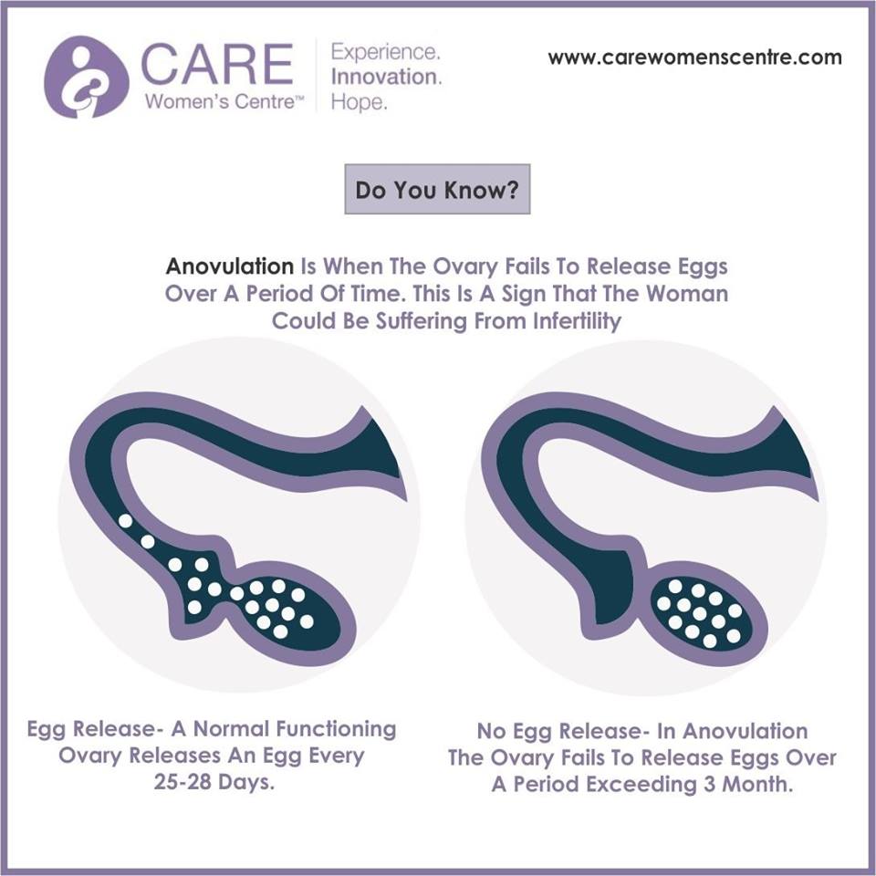
Do you know ? Anovulation is when the ovary fails to release eggs over a period of time. This is a sign that the women could be suffering from infertility